
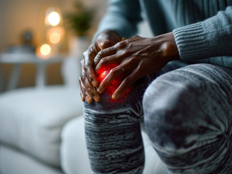
Presentation of bursitis
A bursa (bursae in plural) is a lubricated sac or pocket filled with fluid adjacent to or located between hard or soft tissues that reduces friction and tension between bones, joints, tendons, and/or skin. Bursitis occurs when a bursa becomes irritated or inflamed.
Etiology of bursitis
It can develop in response to trauma, chronic overuse, infection (also known as septic bursitis), or certain systemic rheumatic diseases (e.g., gout, rheumatoid arthritis [RA]). The causes of bursitis are not mutually exclusive (e.g., patients may simultaneously present with septic and gouty bursitis).
Diagnostic evaluation
- Physical examination – The most common signs of bursitis are pain, swelling, and tenderness near a joint.
- Acute bursitis – Pain and functional limitations due to acute bursitis generally develop over days. Acute bursitis of a superficial bursa often causes swelling, warmth, and erythema of the overlying skin, while acute bursitis of a deep bursa is rarely associated with visible changes on examination. Active movement of muscles adjacent to the involved bursa and joint flexion that compresses the bursa generally exacerbate the pain. In contrast, passive movement and joint extension generally do not aggravate the pain unless the affected bursa is compressed.
- Chronic bursitis – Patients with chronic bursitis often present with symptoms that have been present for weeks or even months and may have functional limitations due to pain or contractures and muscle atrophy secondary to immobility. Patients with chronic bursitis of a superficial bursa may present with swelling of the bursa and thickening of the bursal wall that can be palpated on examination. Local warmth and overlying erythema are generally less marked than during an acute process, and the pain is less than expected given the degree of swelling.
- When to aspirate – Aspiration of an inflamed bursa is indicated whenever septic bursitis or bursitis secondary to a crystalline arthropathy is considered. In cases where the affected bursae are deep, relatively inaccessible, and/or close to other important structures (e.g., a major artery or nerve), we use imaging (e.g., ultrasound or computed tomography [CT]) to guide us rather than palpation alone. The bursal fluid should be sent for Gram stain, bacterial culture (aerobic and anaerobic), white blood cell (WBC) count with differential, and crystal evaluation by polarized light microscopy.
- When to perform imaging – Most patients do not require imaging; however, it may be indicated in cases of trauma, when guidance is needed for bursal aspiration or intra-bursal glucocorticoid injection, and/or to exclude other diagnoses when prompt intervention is imperative (e.g., avascular necrosis of the femoral head mimicking iliopsoas bursitis).
Diagnosis
The diagnosis of bursitis is made clinically based on suggestive history and consistent physical examination. The main clinical features depend on the cause, location, and chronicity, but often include tenderness, pain with movements that increase bursal pressure (often joint flexion), swelling of superficial bursae, and joint contractures and/or thickening of the bursal wall in chronic bursitis. Bursal fluid should be non-bloody and bland (leukocytes <500/mm3) in cases of bursitis not caused by infection or gout.
Treatment of bursitis
The primary goal of treating patients with bursitis is to reduce pain and thus improve mobility in the hope of reducing the risk of developing complications such as joint contractures or muscle weakness. It is also important to address any contributing factors, such as excessive joint stress, mechanical imbalance, or underlying systemic rheumatic diseases.
Intra-bursal glucocorticoid injectionsFor most patients with deep bursitis, we suggest an intra-bursal glucocorticoid injection as adjunctive treatment rather than administering NSAIDs alone (Grade 2C). Intra-bursal glucocorticoid injections are not administered in cases of potential septic bursitis risk. Similarly, we generally avoid intra-bursal injections for superficial bursitis due to an increased risk of complications.
Initial therapy for all patientsPatients with all types of bursitis may benefit from conservative management, including relative rest, joint protection, cold compresses, and analgesia. In patients who do not have contraindications to nonsteroidal anti-inflammatory drugs (NSAIDs), we generally offer a one to two-week course of NSAIDs to help reduce inflammation and pain (e.g., ibuprofen 600 to 800 mg three times daily). Complementary therapeutic measures may be useful for patients with certain syndromes (e.g., physiotherapy, specific cushions).
Source: UpToDate